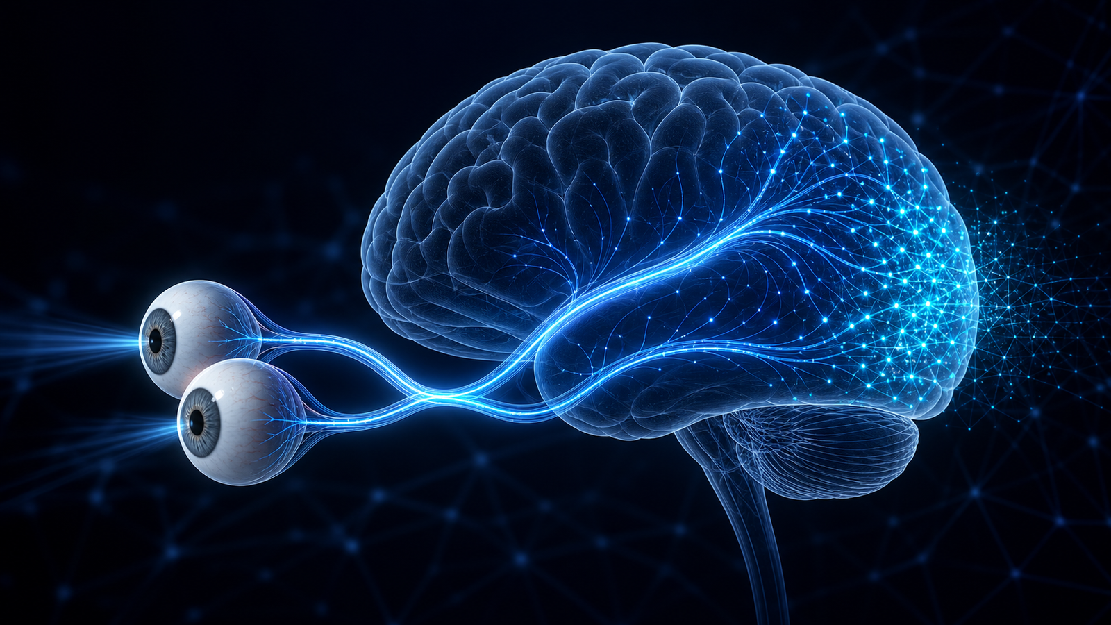
Vision begins when light enters the eyes, but seeing is something the eyes and brain do together. The eyes focus and convert light into neural signals. Networks across the brain organize those signals into shapes, movement, depth, objects, faces, places, and visually guided actions.
Cerebral visual impairment, or CVI, is a spectrum of brain-based visual impairment. A person with CVI may have healthy-looking eyes, an eye condition that does not fully explain their visual difficulty, or both. Understanding CVI starts with understanding that vision is more than eyesight measured on an eye chart.
How vision begins in the eye
The National Eye Institute’s overview of the eye describes the first stages:
- Light is focused. The cornea bends incoming light. The pupil is the opening that lets light enter, while the iris adjusts its size. The lens helps focus light onto the retina.
- The retina converts light into signals. Rod and cone photoreceptors respond to light. Retinal circuits begin processing contrast, colour, detail, and changes over time before the signal leaves the eye.
- The optic nerve carries the output. Axons from retinal ganglion cells form the optic nerve, which carries electrical signals toward the brain.
The retina is neural tissue, not a passive camera sensor. It transforms and organizes information before passing it onward.
From the optic nerve to the visual cortex
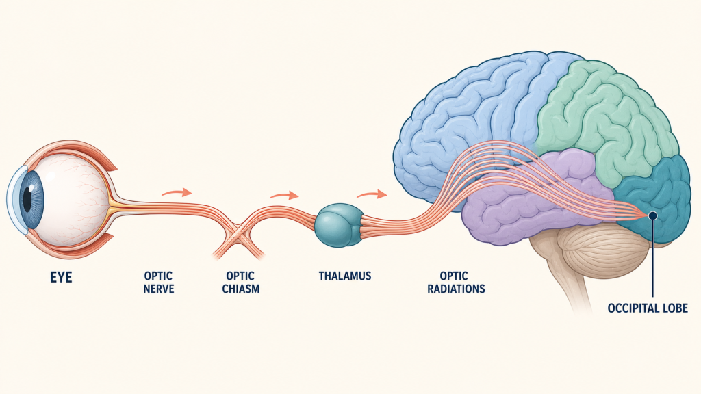
After leaving the eyes, signals travel through a connected pathway described in the NCBI overview of central visual pathways:
- Optic chiasm: some nerve fibres cross to the other side. This arrangement means each cerebral hemisphere receives information about the opposite half of visual space from both eyes.
- Thalamus: much of the signal reaches the lateral geniculate nucleus, a layered relay that preserves spatial organization and sends information onward.
- Optic radiations: these fibre pathways fan through the brain toward the occipital lobe.
- Primary visual cortex: early cortical processing represents features such as edges, orientation, contrast, location, and movement in an organized map of visual space.
- Visual association networks: processing continues across occipital, temporal, and parietal regions that contribute to recognizing what is present and using vision to guide attention and action.
The familiar “what” and “where/how” streams are useful shorthand, not two isolated pipelines. Seeing a crowded room, recognizing a person, judging a step, and reaching for a cup depend on interacting visual, attention, memory, motor, and spatial systems.
Eyes collect and encode visual information; the brain makes that information usable.
What is cerebral visual impairment?
The terms cerebral visual impairment and cortical visual impairment are both used, although “cerebral” reflects that affected pathways can extend beyond the cortex. The 2024 NIH CVI Working Group describes CVI as a spectrum of visual impairments caused by an underlying brain abnormality that affects the development of visual-processing pathways. The visual difficulty is greater than would be expected from any coexisting eye condition alone.
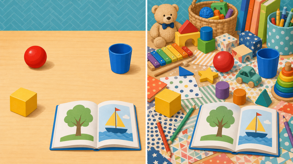
CVI can involve lower-order visual functions, higher-order visual processing, or both. Someone may have reduced acuity or a visual-field difference. Another person may read small letters on an uncluttered chart yet struggle to recognize objects, understand a busy scene, find an item in clutter, process movement, or coordinate looking with reaching.
The definition and terminology are still developing. A systematic review of childhood CVI definitions found substantial variation across research and proposed focusing on verifiable visual dysfunction that cannot be explained by anterior visual-pathway or coexisting ocular disorders.
What CVI may look like
CVI is not one visual experience. The National Eye Institute notes that children with CVI may have difficulty with:
- recognizing faces, objects, or what they are looking at;
- finding something in a visually crowded space;
- understanding busy or moving scenes;
- responding quickly to visual information;
- using vision and reach together; or
- seeing consistently across different environments, levels of fatigue, and competing demands.
These examples are not a diagnostic checklist. The same behaviour can have several explanations, and people with CVI differ widely. Some also have eye conditions, motor differences, communication needs, epilepsy, or other neurological and developmental conditions. CVI is a visual condition; it is not primarily a disorder of language, learning, or social communication.
Why an ordinary eye test may not tell the whole story
Visual acuity is important, but it measures only part of vision under controlled conditions. Functional vision asks how someone uses vision during real activities: finding a person, navigating a room, locating learning materials, interpreting a picture, or reaching safely.
There is no single test that establishes CVI. The American Academy of Pediatrics clinical report recommends a coordinated process that can include medical and developmental history, comprehensive eye examination, observation of visual behaviour, assessment of visual function and functional vision, and—in some cases—neurological evaluation and imaging. An MRI can provide useful information but does not by itself confirm, exclude, or predict an individual’s functional vision.
Support should fit the person and the task
Evaluation can help families, clinicians, educators, and vision professionals identify barriers and test accommodations. Depending on the individual, a team might explore reducing visual clutter, increasing spacing, presenting fewer items at once, using consistent locations, allowing more viewing time, adjusting contrast or lighting, and combining visual information with touch or sound.
These are examples to assess, not a universal CVI programme. Preferences such as a particular colour or moving target are not shared by everyone. A 2023 scoping review of CVI interventions found sparse studies, inconsistent outcome measures, small samples, and generally low-level evidence. Individualized vision services and educational support are established parts of care, while stronger controlled research is still needed to determine which specific interventions work best for whom.
References
- National Eye Institute. How the eyes work. Updated April 20, 2022.
- Purves D, Augustine GJ, Fitzpatrick D, et al., editors. Central visual pathways. Neuroscience. 2nd ed. Sinauer Associates; 2001.
- Chang MY, Merabet LB; CVI Working Group. Cerebral/cortical visual impairment working definition: A report from the National Institutes of Health CVI Workshop. Ophthalmology. 2024;131(12):1359–1365. doi:10.1016/j.ophtha.2024.09.017.
- Lehman SS, Yin L, Chang MY, et al. Diagnosis and care of children with cerebral/cortical visual impairment: Clinical report. Pediatrics. 2024;154(6):e2024068465. doi:10.1542/peds.2024-068465.
- Sakki HEA, Dale NJ, Sargent J, Perez-Roche T, Bowman R. Is there consensus in defining childhood cerebral visual impairment? A systematic review of terminology and definitions. British Journal of Ophthalmology. 2018;102(4):424–432. doi:10.1136/bjophthalmol-2017-310694.
- Delay A, Rice M, Bush E, Harpster K. Interventions for children with cerebral visual impairment: A scoping review. Developmental Medicine & Child Neurology. 2023;65(4):469–478. doi:10.1111/dmcn.15431.
- National Eye Institute. Cerebral visual impairment (CVI). Updated December 4, 2024.
Editorial status: Draft prepared 1 August 2026. Qualified clinical, evidence, editorial, and accessibility review are required before publication. This article provides general education, not diagnosis, medical advice, or an individualized support plan.