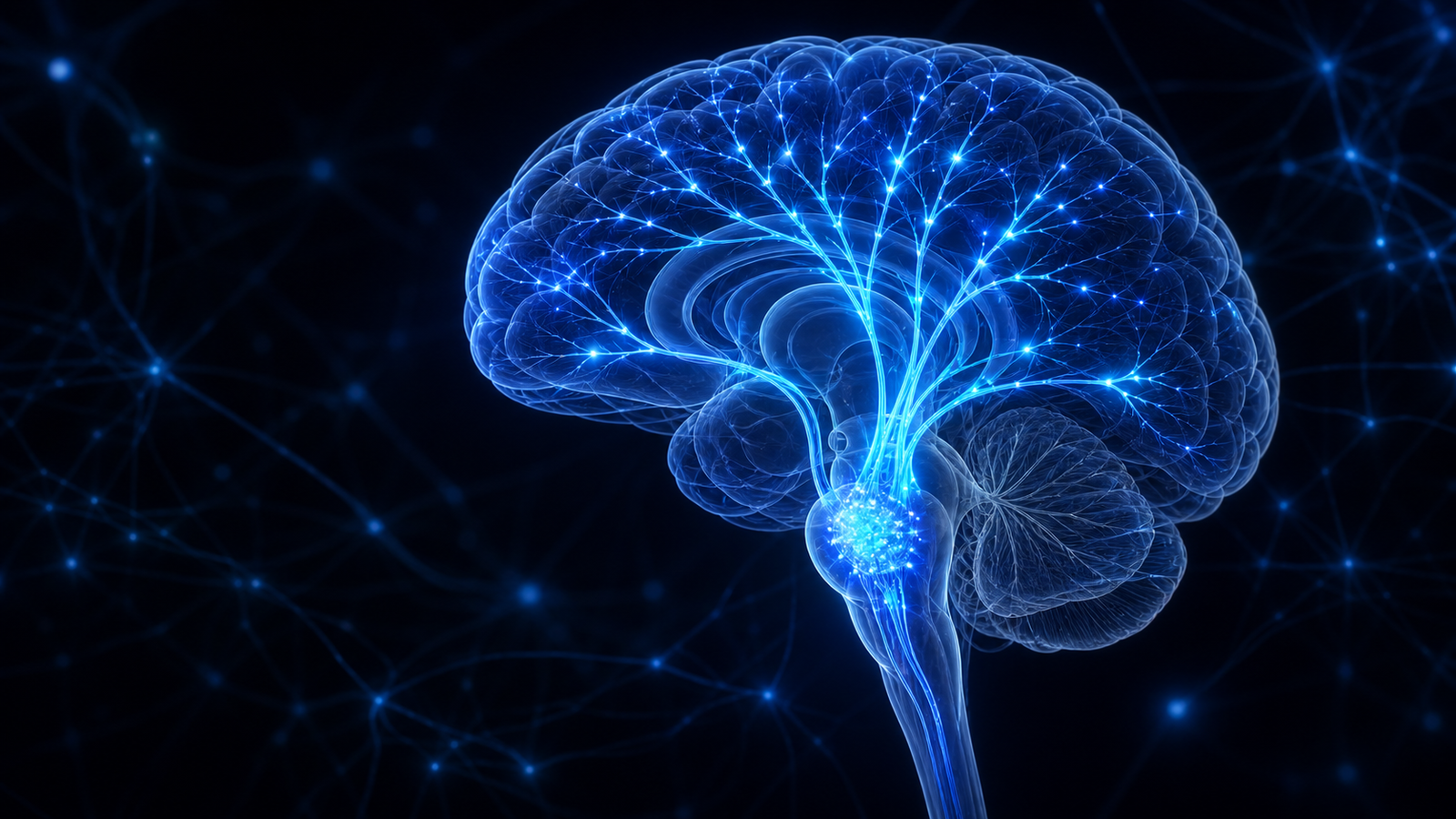
The reticular activating system, often shortened to RAS, is a historical name for brain pathways that help support wakefulness and alertness. It is important—but it is not a single switch, a tiny gatekeeper that chooses every thought, or a mechanism that makes goals appear in the world.
Modern neuroscience describes a distributed ascending arousal system: connected groups of cells in the brainstem, hypothalamus, basal forebrain, and thalamus that influence the cortex and work together with sleep-promoting circuits. Understanding that network helps separate a useful scientific concept from popular claims that stretch the evidence.
RAS in plain language
To respond to a sound, follow a conversation, or notice a signal, the brain first needs an adequate level of wakeful arousal. Ascending arousal pathways help create and maintain that state of readiness by sending widespread signals toward the thalamus, hypothalamus, basal forebrain, and cerebral cortex.
The network uses several signalling systems. Reviews of sleep–wake circuitry describe roles for glutamate and GABA as well as neuromodulators such as acetylcholine, norepinephrine, serotonin, dopamine, histamine, and orexin. These systems overlap and interact; no single chemical or nucleus explains wakefulness by itself.
A useful working description: ascending arousal pathways help make the cortex ready for wakeful, responsive behaviour.
Where the idea came from
In a landmark 1949 animal study, Giuseppe Moruzzi and Horace Magoun stimulated parts of the brainstem reticular formation and observed widespread changes from synchronized electrical activity toward a faster, wake-like pattern in the cortex. This helped establish the idea of an ascending reticular activating system.
The original model was highly influential, but later work revealed a more complicated picture. A review of arousal systems notes that several nuclei surrounding the classical reticular formation are central to widespread cortical modulation. Modern accounts therefore describe multiple interacting pathways rather than one uniform web of neurons.
Human anatomy supports this broader view. A small diffusion-imaging and histology study mapped different brainstem pathways connecting with the thalamus, hypothalamus, and basal forebrain. The study provided proof of principle for a distributed human ascending arousal system; its three-brain sample does not define every pathway or settle how the system works in living people.
Arousal is not the same as attention
Arousal and attention interact, but they are not interchangeable. Arousal describes readiness and responsiveness across states ranging from deep sleep to alert wakefulness. Attention includes processes that prioritize information, orient toward a location or signal, sustain a task goal, and resolve competition.
An influential review of human attention distinguishes alerting, orienting, and executive-control networks. Brainstem arousal systems contribute to alerting, while orienting and executive attention rely on additional cortical and subcortical networks. Being awake enough to respond is necessary for focused attention, but it does not determine by itself what a person selects or how well they resist distraction.
Is the RAS a filter?
Calling the RAS a “filter” can be a useful metaphor only if its limits are clear. The brain cannot process every incoming signal in equal depth, and arousal systems can alter cortical responsiveness. But selection is distributed across sensory pathways, thalamic circuits, memory, expectations, and multiple attention networks. There is no evidence that one RAS switch reviews all information and sends only goal-matching items into awareness.
After choosing a goal—buying a particular bicycle, for example—you may notice related bicycles more often. That experience can reflect top-down attention, memory, expectation, and increased relevance. It does not show that the RAS attracted those bicycles, changed external events, or “manifested” the goal.
What affects alertness?
Alertness changes throughout the day and from one situation to another. Sleep pressure, circadian timing, novelty, motivation, sensory stimulation, medication, health, and the demands of a task can all contribute. These influences act through interacting systems rather than a voluntary RAS on/off control.
Modern reviews describe wake-promoting pathways as overlapping and partly redundant. A 2017 review maps several connected brainstem and hypothalamic systems, while a second wake–sleep circuitry review emphasizes that fast glutamate and GABA signalling forms an important backbone alongside modulatory systems. This is why “activate your RAS” is not a scientifically precise instruction.
A practical interpretation
You do not need to target a particular brain structure to organize attention. More concrete strategies are easier to evaluate:
- Define the next observable action. A specific task gives attention a clearer target than a broad intention.
- Make useful cues visible. Notes, calendars, and environmental prompts reduce reliance on remembering an intention at the right moment.
- Reduce competing signals. Removing avoidable notifications can make selection easier without invoking a special brain hack.
- Check the outcome. Measure whether the strategy changed behaviour instead of treating increased noticing as proof of a neurological explanation.
These are organizational suggestions, not treatments and not claims about changing the RAS. Persistent or concerning changes in alertness require individualized assessment from a qualified health professional; a website or cognitive game cannot determine their cause.
Related reading and practice
Read Sleep: The Brain’s Nightly Reset for a broader introduction to sleep and next-day cognition. The Reaction Time activity involves responding to a signal, but its result is influenced by the device, input method, anticipation, motor response, attention, fatigue, and surroundings. It is not a test of the RAS or of brain health.
References
- Moruzzi G, Magoun HW. Brain stem reticular formation and activation of the EEG. Electroencephalography and Clinical Neurophysiology. 1949;1(4):455–473. doi:10.1016/0013-4694(49)90219-9.
- Scammell TE, Arrigoni E, Lipton JO. Neural circuitry of wakefulness and sleep. Neuron. 2017;93(4):747–765. doi:10.1016/j.neuron.2017.01.014.
- Saper CB, Fuller PM. Wake–sleep circuitry: An overview. Current Opinion in Neurobiology. 2017;44:186–192. doi:10.1016/j.conb.2017.03.021.
- Satpute AB, Kragel PA, Barrett LF, Wager TD, Bianciardi M. Deconstructing arousal into wakeful, autonomic and affective varieties. Neuroscience Letters. 2019;693:19–28. doi:10.1016/j.neulet.2018.01.042.
- Petersen SE, Posner MI. The attention system of the human brain: 20 years after. Annual Review of Neuroscience. 2012;35:73–89. doi:10.1146/annurev-neuro-062111-150525.
- Edlow BL, Takahashi E, Wu O, et al. Neuroanatomic connectivity of the human ascending arousal system critical to consciousness and its disorders. Journal of Neuropathology & Experimental Neurology. 2012;71(6):531–546. doi:10.1097/NEN.0b013e3182588293.
Editorial status: Draft prepared 1 August 2026. Evidence, editorial, and accessibility approval are required before publication. This article provides general education, not medical advice or cognitive assessment.